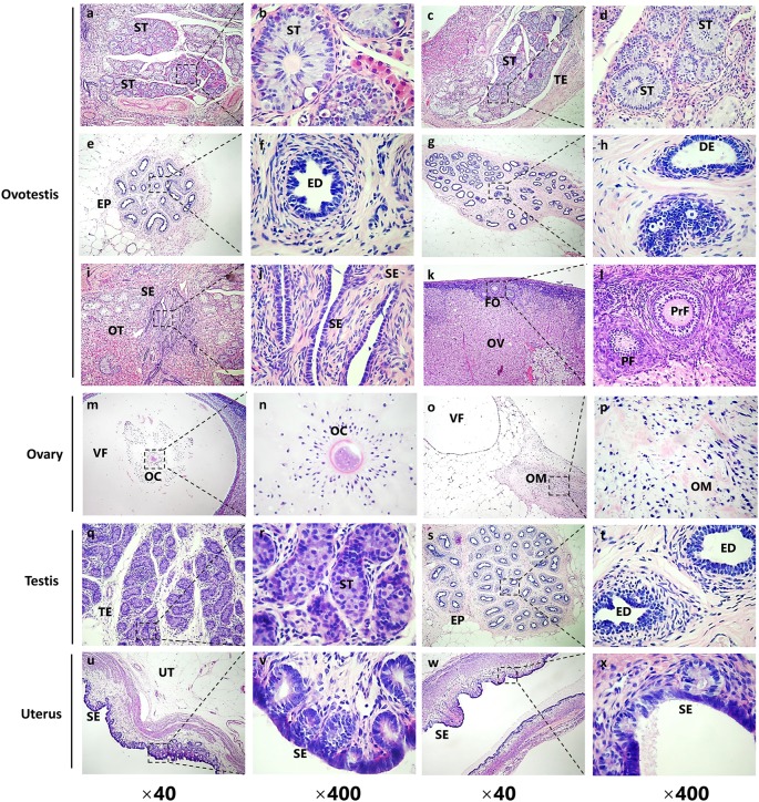
Figure 3. Histological examination of the gonadal structures by H&E staining from SRY-mutant chimeric rabbits.
(a–l) H&E staining of the left gonad (ovotestis in Figure 2) from the SRY-mutant chimeric rabbit. (a–h) Male-like gonadal structures of the ovotestis. ST, seminiferous tubules; TE, testis; EP, epididymis; ED, efferent ductules; DE, ductus epididymidis. (i-l) Female-like gonadal structures of the ovotestis. SE, surface epithelium of the endometrium; OT, ovotestis; FO, follicles; OV, ovary; PF, primordial follicles; PrF, primary follicles. (m–p) H&E staining of the upper right gonad (ovary in Figure 2) from the SRY-mutant chimeric rabbit. VF, vesicular follicles; OC, occytes; OM, ovary medulla. (q–t) H&E staining of the bottom right gonad (testis in Figure 2) from the SRY-mutant chimeric rabbit. ST, seminiferous tubules; TE, testis; EP, epididymis; ED, efferent ductules; (u–x) H&E staining of the uterus from the SRY-mutant chimeric rabbit. UT, uterus; SE, surface epithelium of the endometrium.

