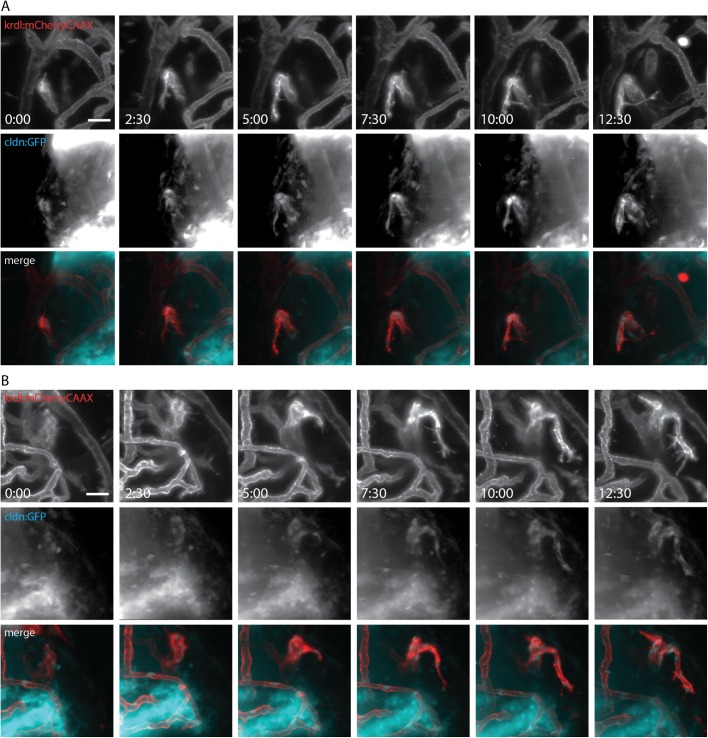
Fig. 4.
Initiation of claudin 5a expression coincides with blood vessel formation. (A,B) Still images of two time lapses of blood vessel development in Tg(kdrl:mCherry)is5;TgBAC(cldn5a:EGFP)vum2 larva at 96 hpf. Images were taken every 2.5 h for a period of 12.5 h on blood vessels in the optic tectum of the midbrain. Images of single channels show blood vessel development (kdrl:mcherryCAAX) on the top row, coinciding with development of claudin 5a in the middle row. Merged images are shown in the bottom row. Scale bars: 20 μm. See corresponding movies of time lapses (Movies 1 and 2).

