Fig. 5.
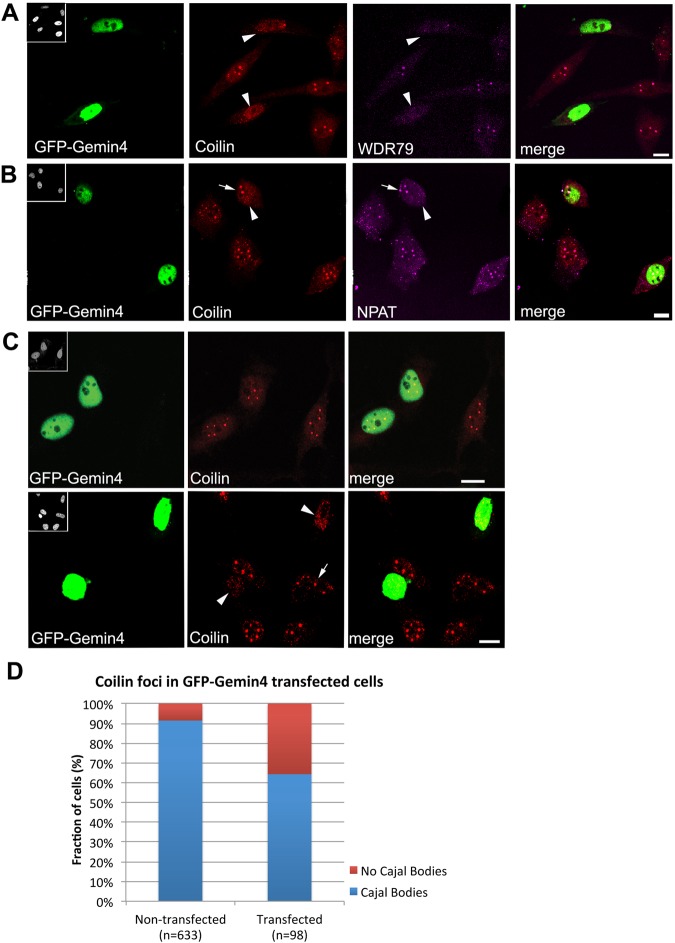
Cajal bodies are disrupted by high levels of GFP-Gemin4 expression. HeLa cells were transfected with GFP-Gemin4 (shown in green) and then co-stained (in red) with antibodies targeting coilin (A-C), WDR79 (A) and NPAT (B). Insets in upper left corners of GFP images show the DAPI-stained nuclei. Arrowheads indicate transfected cells. Arrows indicate coilin foci corresponding to Cajal bodies. Cells strongly expressing GFP-Gemin4 show major disruptions to Cajal bodies, whereas lower expression levels of GFP-Gemin4 had little effect on cells (B). Nuclei were scored for the presence or absence of coilin foci (D). Chi squared analysis reveals a significant difference between the two cell populations, P<0.001. Scale bars: 10 µm.

