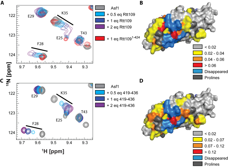
Figure 1.
Rtt109 interacts with Asf1 via its C-terminus. (A) Overlay of 15N-HSQC spectra of Asf1 (100 μM) in the presence of Rtt109 and Rtt1091–424. The target concentrations of full-length Rtt109 corresponded to 0.5, 1 and 2 equivalents of Rtt109, but the actual achieved concentrations (estimated from 1D 1H spectra) were lower due to partial precipitation of Rtt109 in the NMR tube. The spectra were acquired in 50 mM sodium citrate pH 6.5, 150 mM NaCl, 5 mM BME at 600 MHz and 298 K. (B) Mapping of the CSPs observed upon addition of 1 equivalent of full-length Rtt109 onto the Asf1 structure in complex with the H3:H4 dimer (PDB ID 2HUE). (C) Overlay of 15N-HSQC spectra of Asf1 (100 μM) in the presence of Rtt109419–436. The spectra were acquired in 50 mM sodium citrate pH 6.5, 150 mM NaCl, 5 mM BME at 850 MHz and 298 K. (D) Mapping of the CSPs observed upon addition of 1 equivalent of Rtt109419–436 onto the Asf1 structure as in (B). The chemical shifts perturbations observed upon addition of Rtt109419–436 were larger than those observed upon addition of full-length Rtt109, which can be recapitulated by the on-going precipitation of full-length Rtt109 during the NMR experiment. Asf1 is in surface representation.

