Fig. 10.

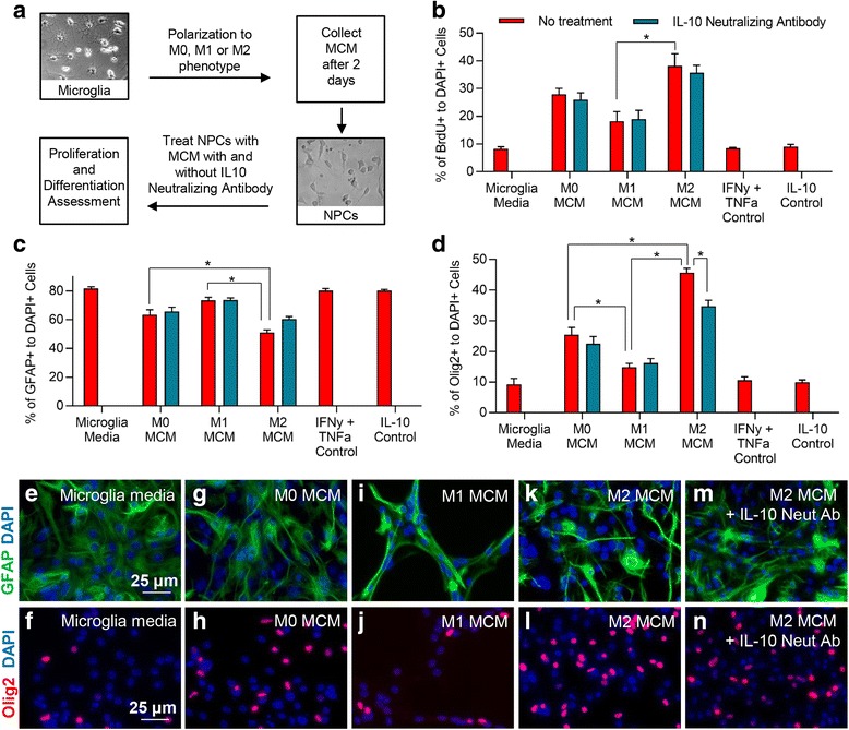
M2 microglia promote oligodendrocyte differentiation of spinal cord NPCs through IL-10. a To assess the effects of microglia effects on NPC proliferation and differentiation, MCM was collected from microglia 2 days following polarization. This media was then transferred to NPC cultures to assess proliferation and differentiation. b MCM derived from M2 microglia significantly promoted NPC proliferation compared to M1 MCM. IL-10 neutralizing antibody had no effect on the overall proliferation of NPCs by M2 MCM suggesting this effect was not mediated through IL-10. c A significant decrease was observed in the percentage of GFAP+/DAPI+ astrocytes when NPCs were exposed to M2 MCM (k) compared to both M0 (g) and M1 (i) MCM. (d) M2 MCM (l) significantly increased the percentage of Olig2+/DAPI+ cells compared to both M0 (h) and M1 (j) MCM. Additionally, M1 MCM significantly reduced the percentage of Olig2+ cells compared to both M0 and M2 MCM. m, n The effect of M2 MCM was significantly reduced by IL-10 neutralizing antibody. e, f represents NPCs N = 3–5 independent experiments. The data show the mean ± SEM, *p < 0.05, one-way ANOVA
