Fig. 7.

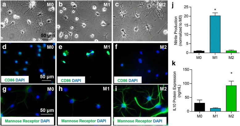
Polarization of primary microglia cultures to an M0, M1, or M2 phenotype. a–c Primary microglia were polarized to M1 through IFNγ and TNFα treatment or M2 through IL-10 treatment. M1 polarization was confirmed by induced expression of CD86 (d–f) and release of nitrite (j). Increased expression of mannose receptor (g–i) and IL-10 (k) were used to confirm M2 polarization. N = 5 independent experiments. The data show the mean ± SEM, *p < 0.05, one-way ANOVA
