
Figure 4.
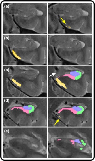
Illustration of ASHS segmentation errors in the initial, nonoptimized validation attempt. Left column depicts correct, manual segmentation, and right column shows faulty segmentations. (a) Whereas manual segmentation (left) does not include this slice in the range, ASHS (right) includes multiple, erroneously included voxels in ERC, as indicated by the yellow arrow. (b) Manual segmentation of ERC (left) in comparison with omitted segmentation by ASHS (right). (c) Manual segmentation includes only ERC (left) as visible presence of uncus (indicated by the white arrow) indicates no body segmentation on this slice. In contrast, ASHS (right) includes segmentation of ERC and body subregions. (d) Overextension of ERC by ASHS in several voxels (right, as indicated by the yellow arrow), where ERC should no longer be segmented following the first body slice (left). (e) Following the disappearance of the lamina quadrigemina, subfields are no longer segmented by the manual approach (left), but are both included, and mis‐segmented by ASHS (right) [Color figure can be viewed at http://wileyonlinelibrary.com]