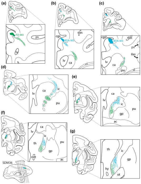
FIGURE 11.
Line drawings of representative rostral (a) to caudal (g) coronal sections from case SDM36 depicting the descending course of labeled axon pathways in the CR and IC following injections of tract tracer into M3. Specifically, FD was injected into the central part of the orofacial region and BDA was injected into the caudal part of the orofacial area and rostral part of the M3 arm area (see Figure 8, bottom). The green fiber bundles represent axons originating from the FD injection site and the blue fiber bundles represent axons from the BDA injection site. Note how the respective fiber bundles arch around the head of the caudate (ca), enter the internal capsule (IC), and remain relatively spatially separate throughout the descending route, with a label sparse zone in between that contains a light mixture of FD and BDA labeled fibers. For other abbreviations see list

