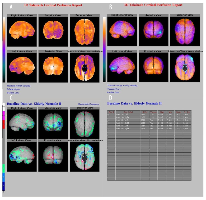
Figure 3.
A 48-years-old male patient. (A, B). Z-value map of semi-quantitative analysis by NeuroGam software. (C) Comparison of rCBF hypoperfusion area in Brodmann 10, 45, 46 zones of the left frontal lobe and normal databases (represented by standard deviation). (D). The left frontal Brodmann 10, 45, and 46 zones were decreased by 2.4, 3.9, and 3.1 SD, respectively, and the right frontal Brodmann 10, 45, and 46 zones were decreased by 1.3, 1.2, and 1.0 SD, respectively, suggesting that the lesion was located in the left frontal lobe (Brodmann 10, 45, 46 zone). rCBF – regional cerebral blood flow.

