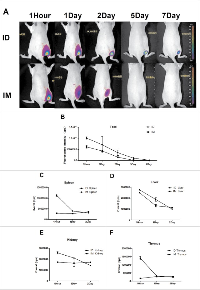
Figure 4.

The H7N9 split vaccine was labeled with Cy5 for visualization of antigen deposition and metabolism in vivo. Rats were immunized ID using MP-0.1 or IM using a syringe and anesthetized with 4% isoflurane. Fluorescence signals were visualized at 1 hour, 1 day, 2 days, 5 days, and 7 days, and background auto-fluorescence was subtracted (A). Fluorescence intensity over time (B). At 1, 24, and 48 h, three rats per group were euthanized, and the kidney, liver, spleen, and thymus were removed and subjected to fluorescence imaging (C, D, E, F).
