
FIG 10.
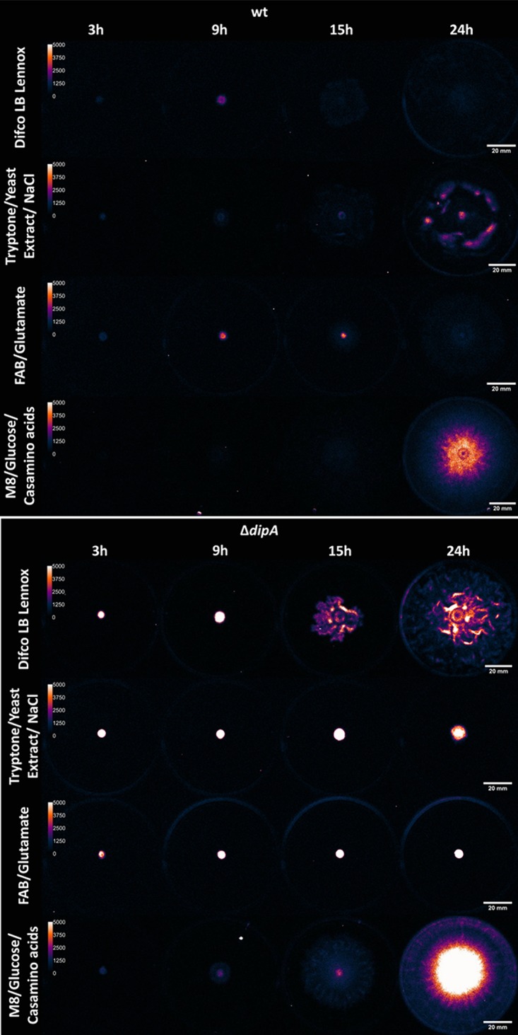
In vivo luminescence images show the variation of c-di-GMP throughout the swarming event on different media. After a 3-h incubation, the c-di-GMP PcdrA::lux reporter emits a weaker signal in the wt (top) than in the ΔdipA mutant (bottom), as would be expected when a phosphodiesterase is removed. There is no significant difference between the reporter signals in the wt on the media tested. Contrastingly, there is differential reporter activity in the ΔdipA mutant, further indicating that the role of DipA is nutritionally conditional. As would be expected, the nonswarming ΔdipA mutant on tryptone, yeast extract, and NaCl emits a very high signal compared to when it swarms on LB Lennox broth. However, the most striking result is the robust swarming combined with high levels of c-di-GMP seen when cultivated on M8 medium with glucose and Casamino Acids. Each plate has a 2-μl drop of broth, normalized to an OD600 of ≈0.5 before inoculation and incubation. Pictures were taken after 3, 9, 15, and 24 h of incubation at 30°C. Brightness maximum was set to 5,000 photons/s/mm2. Luminescence images are falsely colored for contrast.