Figure 2.
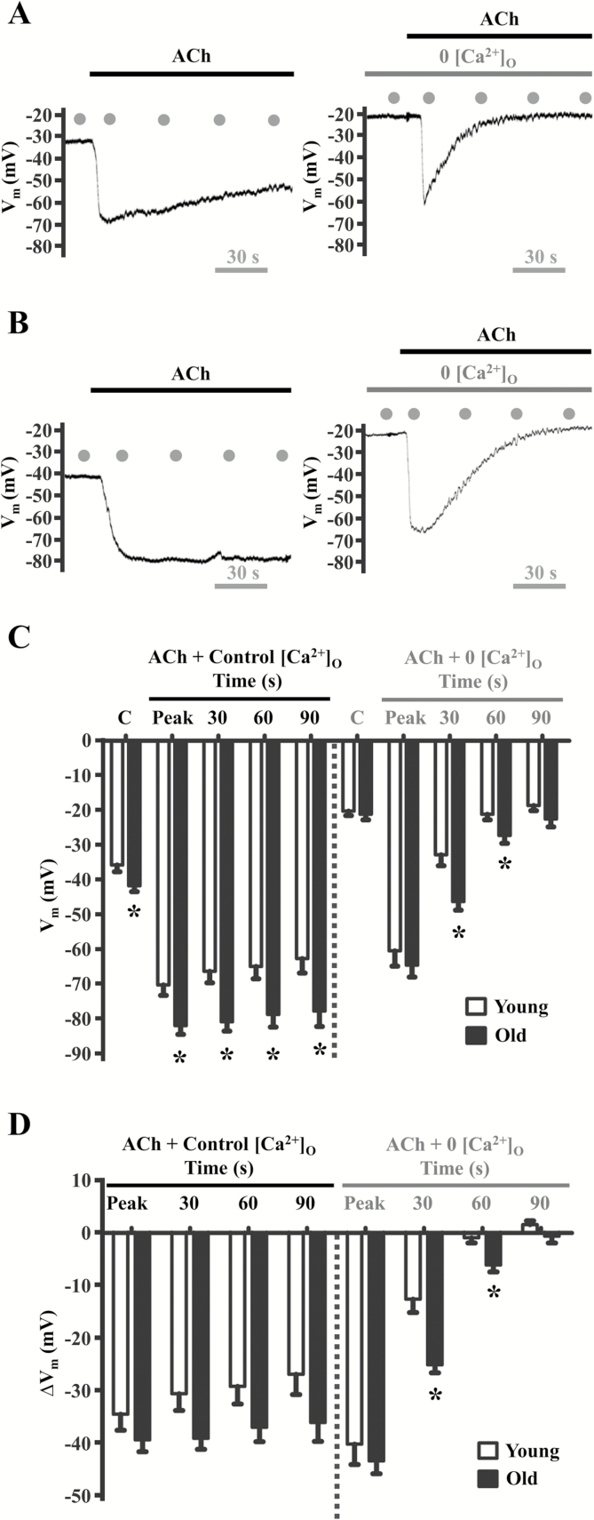
Aging enhances hyperpolarization in response to ACh. Vm recordings (obtained simultaneously with Fura-2 data in Figure 2) in response to 3 × 10⁻⁶ M ACh in the presence of 2 × 10⁻³ M [Ca2+]o (Control, “C”) and with 0 [Ca2+]o in A, Young and B, Old endothelium. Summary data indicate C, Vm and D, ΔVm (treatment − control) for respective age groups. Note initial “peak” of hyperpolarization followed by sustained “plateau” through 90 s (A and B, left panels). Defined time points indicated by gray dots in panels A and B. Note diminished plateau during 0 [Ca2+]o to eliminate Ca2+ entry (A and B, right vs. left panels)]. Peak Vm responses were greater (p < .05) in old vs. young in the presence of 2 × 10⁻³ M [Ca2+]o (panels C and D). Further, Vm remained hyperpolarized (*p < .05) in old vs. young at 30 s and 60 s during 0 [Ca2+]o, reflecting sustained internal release of Ca2+ (panel D). Traces for Figures 1A and 2A (Young) and Figures 1B and 2B (Old) represent simultaneous [Ca2+]i and electrical measurements. *p < .05, old vs. young, n = 10 per group. ACh = acetylcholine.

