Fig. 3.
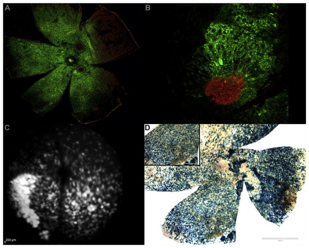
AAV2 successfully transduces subretinal and outer retinal tissues. A. RPE/choroid from an eye treated with AAV2.AcGFP (green) and stained with isolectin (red). Red neovascular tufts (at 2 and 5 o’clock) surrounded by diffuse GFP signal validate CNV formation within the AAV2 treated tissue. B. High power magnification of laser-induced CNV (isolectin, red) within AAV2.AcGFP treated RPE/choroid (green). C. Funduscopic FA modality image showing diffuse in vivo expression of GFP autofluorescence in an eye AAV2.AcGFP. D. RPE/choroid from an eye treated with AAV2.LacZ and stained with X-gal. Blue signal indicates successful AAV transduction following subretinal administration. Scale bars = 1000 μm main, 400 μm inset. (For interpretation of the references to colour in this figure legend, the reader is referred to the web version of this article.)

