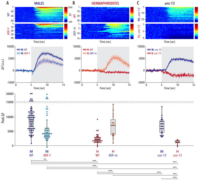
Figure 3. The sexual state of ADF governs its ability to respond to ascaroside stimulation.
Upper panels show pseudocolored graphs of calcium recordings of individual neuronal responses to stimulation with an ascr#2/#3/#8 mixture. Each animal underwent three rounds of stimulation; each row depicts a single response, sorted by peak activity. The pseudocolor scale represents GCaMP3 ΔF (red highest, blue lowest). Middle panels show average ΔF intensities ± SEM for the recordings depicted in the upper panels. Gray shading indicates the period of ascaroside stimulation. Lower panels show peak ΔF values for each recording; dots represent individual responses to each pulse. (A) Wild-type males showed robust calcium transients in ADF upon ascaroside stimulation. The strength of this response was significantly reduced in ADF-feminized males. The modest reduction in mean response (central panel) likely reflects a bimodal distribution of responses arising from variable effects of the ADF feminization transgene, more clearly apparent in the lower panel. (B) In wild-type hermaphrodites, ADF appeared insensitive to ascaroside stimulation. Masculinization of ADF in an otherwise wild-type hermaphrodite was sufficient to confer robust ascaroside sensitivity to ADF. (C) unc-13 mutants maintained a marked sex difference in the response of ADF to ascaroside stimulation. n = 9–66 trials (3–22 animals with 3 trials per animal). Groupwise comparison of peak ΔF values was carried out using Kruskal-Wallis analysis and Dunn’s posthoc test with Bonferroni correction. **p ≤ 0.01, ***p ≤ 0.001. See also Figure S3.

