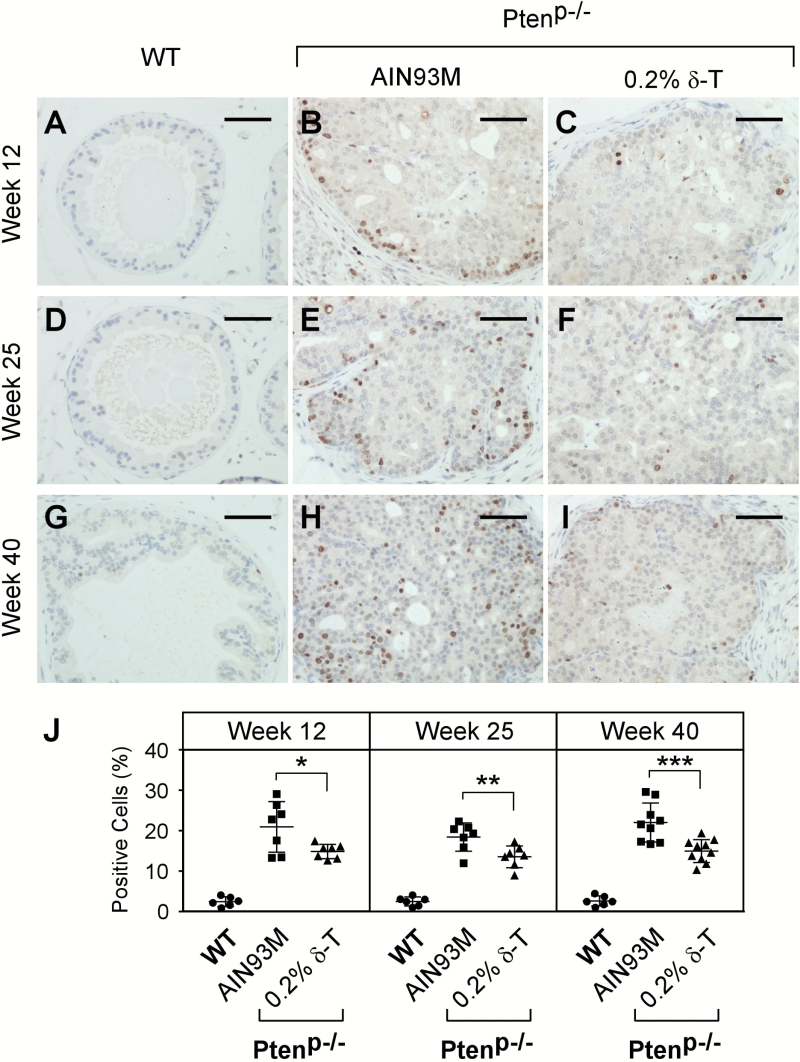
Figure 5.
Dietary δ-T reduced cell proliferation in prostate of Ptenp−/− mice. Prostates of the WT mice fed an AIN93M and Ptenp−/− mice fed either an AIN93M or a 0.2% δ-T diet were analyzed for cellular proliferation using IHC stainingfor Ki67. Images of representative IHC staining for the mouse prostate samples at the ages of 12, 25 and 40 weeks are shown (A–I). The scale bar represents 50 µm. The quantified results of positive Ki67 stained cells in the three groups of mice were determined using the Aperio ScanScope and are summarized in (J). Data are presented as mean ± SD (Experimental conditions are the same as Figure 4). *P = 0.029, **P = 0.013 and ***P = 0.01.

