Figure 1.
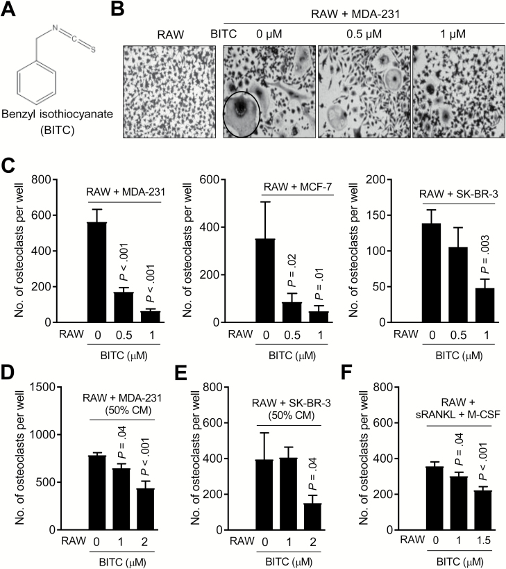
BITC treatment inhibits human breast cancer-induced osteoclast differentiation in vitro. (A) Chemical structure of BITC. (B) Representative microscopic images (×200 magnification) depicting osteoclasts (circled) in co-cultures of RAW264.7 (RAW) and MDA-MB-231 (MDA-231) cells after treatment with DMSO or the indicated doses of BITC for 6 days. Osteoclast differentiation was not observed in RAW264.7 cells alone. (C) Quantitation of osteoclast number in co-cultures of RAW264.7 (RAW) and specified breast cancer cells after treatment with DMSO or the indicated doses of BITC for 6 days. Quantitation of osteoclast number in RAW264.7 (RAW) cultured for 6 days with CM from MDA-MB-231 (MDA-231) (D) or SK-BR-3 cells (E) with or without BITC treatment. (F) Effect of BITC treatment on osteoclast number in RAW264.7 (RAW) stimulated by addition of sRANKL (100 ng/ml) and M-CSF (30 ng/ml) for 6 days. Columns indicate average of triplicate samples from a representative experiment, and bars indicate standard deviation. P value was determined by one-way analysis of variance with Dunnett’s adjustment. Consistent results were obtained in at least two independent experiments.

