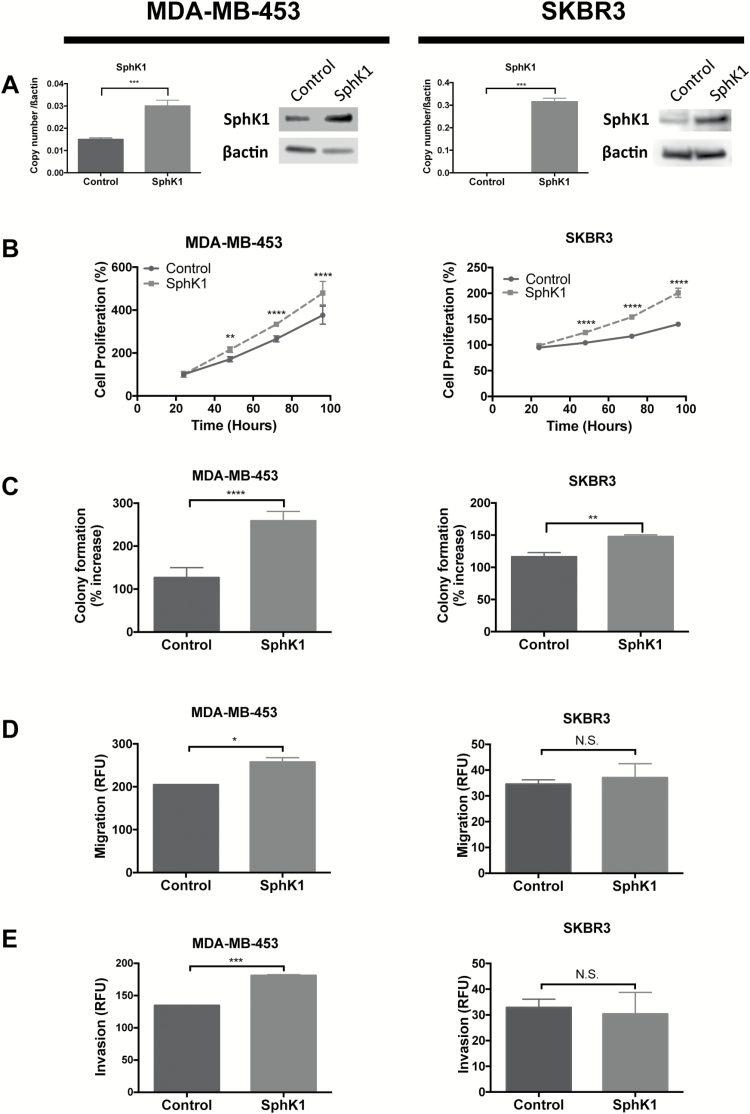
Figure 3.
SphK1 overexpression increased cell growth and aggressiveness MDA-MB-453 and SKBR3 cell lines. MDA-MB-453 and SKBR3 cells transfected with SphK1 and stable cell lines overexpressing SphK1 were generated. (A) qRT-PCR and SphK1 of SphK1 genes in MDA-MB-453 and MDA-MB-231 cells stably transfected with SphK1 or mock control plasmid. (B) Cell proliferation monitored by MTT assay. Cells were seeded at 2.5 × 10³ cells or 5 × 10³ cells and proliferation was measured at 24, 48, 72 and 96 h. (C) Colony formation measured by soft agar assay. Cells were seeded at 5 × 10³ cells and colony formation was measured after 6–10 days. (D) and (E) Migration and invasion assays. Serum starved cells were seeded at 0.25 × 10⁶ to 0.5 × 10⁶ cells in polycarbonate membrane (D) or matrigel coat (E) inserts and allowed to migrate/invade against 10% serum. (A)–(E) Data from one representative experiment are presented as mean ± SEM; *P < 0.05; **P < 0.01; ***P < 0.001 and ***P < 0.001.

