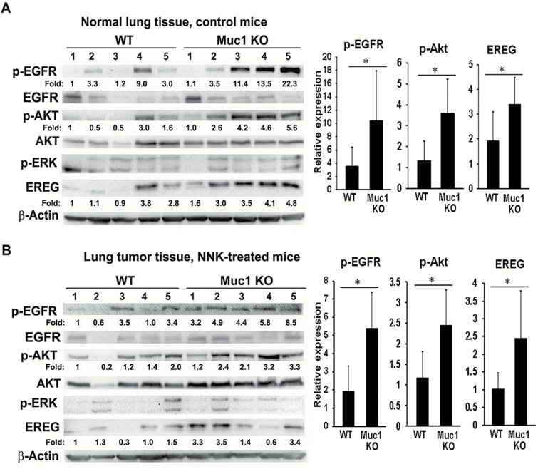
Figure 2.
The EGFR/Akt pathway was activated in both the lung and tumor tissues in the Muc1 KO mice. Normal lung tissue (A) and tumor tissues (B) from five randomly selected mice in both the WT mice group and the Muc1 KO group were subjected to Western blot assay. The expression of phospho-EGFR (Y1068), -Akt (Ser 473), and -ERK (Y185/187) was determined. The expression of EREG, total EGFR, and Akt was also detected by Western blot. β-Actin was detected as an input control. The intensity of the individual bands was quantified and normalized to the corresponding input control bands. Relative expression of each protein was statistically analyzed and shown to the right of its respective panel. *P < 0.05.

