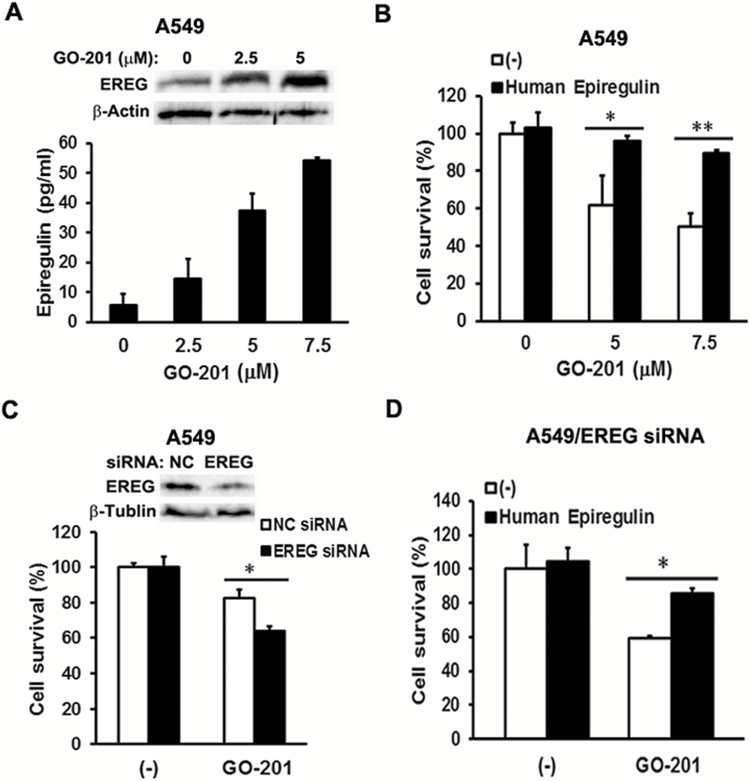
Figure 4.
EREG protects human lung cancer cells from MUC1 inhibitor GO-201 induced cell death. (A) Upper, A549 cells were treated with MUC1 inhibitor GO-201 (0, 2.5 and 5 µM) for 24 h. EREG expression was detected by Western blot. β-Actin was detected as an input control. Lower, A549 cells were cultured in 0.5% FBS containing RPMI medium overnight, before the cells were treated with GO-201 (0, 2.5, 5 and 7.5 µM) for 8 h. After treatment, conditioned media were collected for detection of EREG production by ELISA assay. (B) A549 cells were cultured in 0.5% FBS containing RPMI medium overnight, before the cells were exposed to human EREG (20 ng/ml) for 24 h. After EREG treatment, the cells were treated with GO-201 (0, 5 and 7.5 µM) for 48 h. Cell viability was detected by MTT assay. Data shown are mean ± S.D; **P < 0.01, *P < 0.05. (C) A549 cells were cultured in 10% FBS containing RPMI medium overnight, before the cells were transfected with EREG siRNA or negative control siRNA for 24 h. The cells were then maintained in 0.5% FBS containing RPMI medium for another 24 h. After that, the A549/NC siRNA and A549/EREG siRNA cells were treated with GO-201 (5 µM) for 48 h. Cell viability was detected by MTT assay. Data shown are mean ± S.D; *P < 0.05. (D) The A549/EREG siRNA cells were pre-treated with human EREG (20 ng/ml) for 24 h, before they were treated with GO-201 (5 µM) for 48 h. Cell viability was detected by MTT assay. Data shown are mean ± S.D; *P < 0.05.

