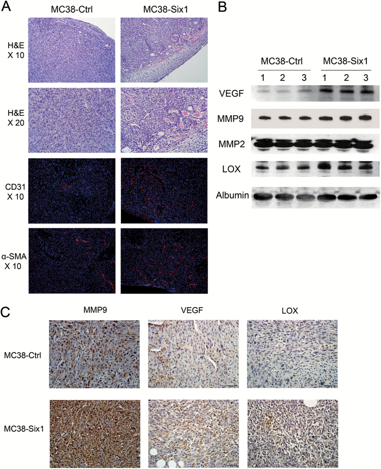
Figure 3.
Six1 overexpression promotes angiogenesis. (A) Upper panels: H&E staining of primary tumors formed in the cecum of mice from MC38-Ctrl and MC38-Six1. Lower panels: immunofluorescent staining of CD31 (red) and α-SMA (red) merged with nuclei (blue) in MC38-Ctrl and MC38-Six1 tumors. (B) Western blots for VEGF, MMP9, MMP2 and LOX in sera from MC38-Six1 tumor-bearing mice and MC38-Ctrl mice. Albumin was used as a loading control. (C) The expression of MMP9, VEGF and LOX in MC38-Six1 and MC38-Ctrl tumors as assessed by immunohistochemistry (×400).

