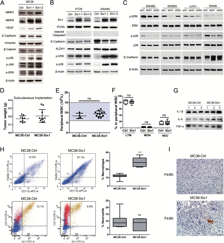
Figure 5.
(A–C) Six1 actives MAPK in vitro. (A) Western blots for MMP2, MMP9, VEGF, E-cadherin, vimentin, β-catenin, p-p38, p-JNK and p-ERK in two different isolates of MC38-Six1 and a MC38-Ctrl line. (B) Western blots for Six1, PCNA, cleaved caspase-3, E-cadherin, ALDH1, p-ERK and p-p38 in two different isolates of Six1-overexpressing HT29 (HT29-Six1) and SW480 (SW480-Six1) and corresponding controls (HT29-Ctrl and SW480-Ctrl). (C) Western blots for p-ERK, ERK, p-p38, p38 and E-cadherin in Six1-knockdown SW480 (SW480-shS1 and SW480-shS2), SW620 (SW620-shS1 and SW620-shS2), LoVo (LoVo-shS1 and LoVo-shS2) and SW48 (SW48-shS1 and SW48-shS2) as well as corresponding controls (SW480-shC, SW620-shC, LoVo-shC and SW48-shC). β-Actin was used as a loading control. (D–I) MC38 cells overexpressing Six1 recruit macrophages to tumors. (D) MC38-Six1 or MC38-Ctrl were injected into the flank of C57BL/6 mice and the mice killed 6 weeks later (n = 9–10). The weight of tumors formed from MC38-Ctrl and MC38-Six1 is shown. (E) Peripheral white blood cell (WBC) count from MC38-Ctrl and MC38-Six1 tumor-bearing mice (cecal implantation). (F) Percentage of lymphocytes (LYM), monocytes (MON) and neutrophils (NEU) in peripheral WBC from MC38-Ctrl and MC38-Six1 tumor-bearing mice (cecal implantation). (G) Western blot of IL-1β, IL-6 and TNF-α in sera from MC38-Six1or MC38-Ctrl tumor-bearing mice (cecal implantation). (H) Left panels: Flow cytometry analysis of the expression of CD11b, F4/80 and Gr1 in MC38-Ctrl and MC38-Six1 tumors from the cecum. Right panels: the percentage of total cells positive for either CD11b+/F4/80+ (a marker for macrophages) or CD11b+/Gr1+ (a marker for neutrophils) cells. Results are representative of seven independent experiments. Bars indicate SD and *** indicates P < 0.001. (I) Immunohistochemical analysis of the expression of F4/80 in MC38-Ctrl and MC38-Six1 derived tumors (×400).

