Figure 3.
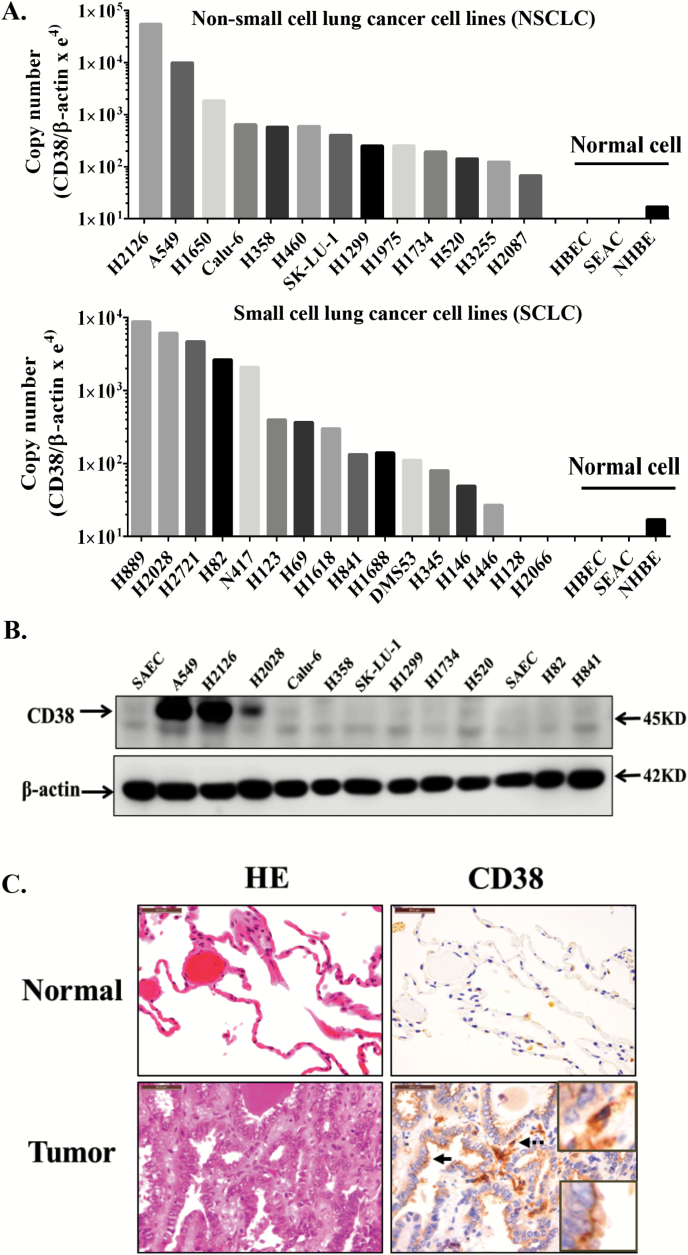
CD38 expression in human lung cancer samples and cell lines. (A) Quantitative real-time RT-PCR analysis of CD38 mRNA expression in multiple non-small and small cell lung cancer cell lines. (B) Immunoblotting of CD38 in lung cancer cell lines. (C) Representative HE staining images show the typical morphology of lung adenocarcinoma and adjacent normal tissue. Serial sections from the same sample were used for immunohistochemically analysis of CD38. CD38-positive lymphocytes/macrophages (dotted arrow) were seen in all sections, with enlarged images in upper-right square. CD38-positive tumor cells (solid arrow) were detected only in some samples, enlarged images in bottom-right square. Scale bar: 50 μm. Experiment was repeated three times with similar results. HBEC, human bronchial epithelial cells; NHBE, normal human bronchial epithelial cells; SEAC, human small airway epithelial cells.

