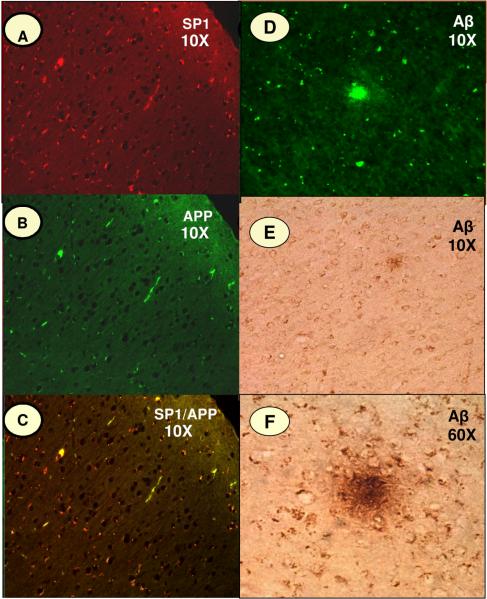
Fig. 6. SP1, APP and Aβ Immunoreactivity in Monkey Cortex.
Intra-neuronal staining of SP1, APP and Aβ in the frontal association cortex of cynomolgus monkey (Macaca fascicularis). The fluorescence immunoreactivity of (A) SP1, (B) APP, and (D) Aβ; (C) shows the co-localization of SP1 and APP; (E & F) reveals the staining patterns of Aβ in senile plaques.

