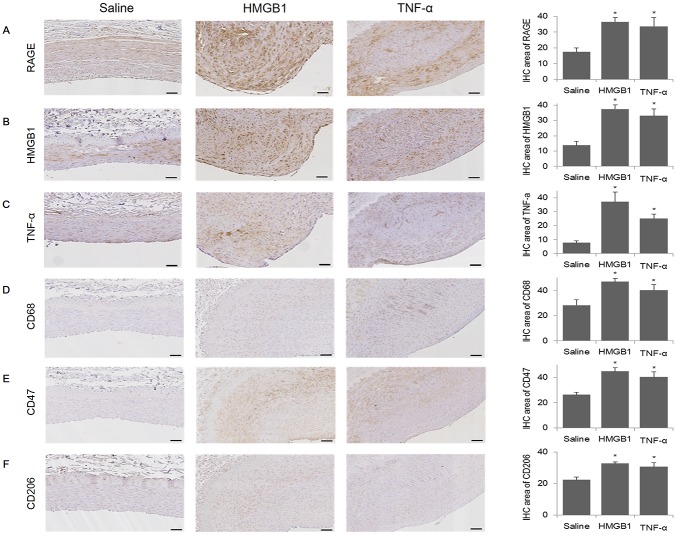
Fig 2. IHC analysis of the degree of inflammation and macrophages present in the arteries of mini-pigs with induced atherosclerosis.
Inflammation and macrophages present in arterial plaques was detected by Immunohistochemical (IHC) staining for RAGE, HMGB1, TNF-α, CD68, CD47, or CD206 in the saline (n = 11), HMGB1 (n = 11), and TNF-α (n = 10) groups. Representative images of IHC staining of RAGE (A), HMGB1 (B), TNF-α (C), CD68 (D), CD47 (E), and CD206 (F) in the mini-pig artery (amplification 100×). IHC area was calculated as (intima/plaque) × 100, %. Scale bars represent 100 μm. Quantitative data are represented as the mean±SEM. *p<0.05, compared with the saline group.

