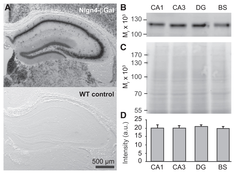
Figure 1. Nlgn4 Expression in the Hippocampus.
(A) (Top) Assessment of Nlgn4 promoter activity using the β-galactosidase reporter expressed by the gene trap in the Nlgn4 KO line (Jamain et al., 2008) and X-gal staining reveal expression of the reporter throughout the hippocampus. (Bottom) WT control shows a lack of X-gal staining in the absence of β-galactosidase.
(B–D) Nlgn4 levels in WT hippocampal CA1 and CA3 regions, dentate gyrus (DG), and brainstem (BS). Representative Nlgn4 immunoblot (B), corresponding protein loading (C, Memcode stain), and quantification (D) are shown (n = 5 animals).

