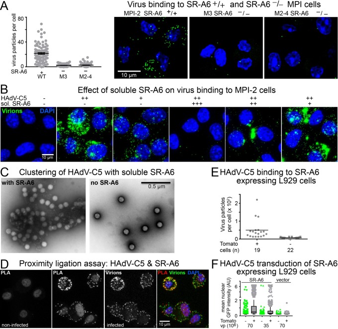
Fig 2. SR-A6 facilitates virus binding to MPI-2 cells.
A) Binding of Alexa-Fluor488-labeled HAdV-C5 to parental MPI-2 cells or the mSR-A6-/- M3 and M2-4 cell lines. Virus was added to cells at +4°C for 60 min (moi ~ 2540 virus particles per cell) and cells were shifted to 37°C for 5min before analysis. The plot shows number of bound virus particles per cell, one dot representing one cell. Error bars represent the means ± SEMs. The difference between SR-A6-positive and SR-A6 knockout cells was statistically significant (P<0.0001, Kolmogorov-Smirnov test). The right-hand panel shows representative images from the three cell lines analyzed. Images are maximum projections of confocal stacks. Virus particles are shown in green and nuclei (DAPI) in blue. Scale bar = 10 μm. B) Effect of preincubation of HAdV-C5 with three different amounts of soluble extracellular domain of mouse SR-A6. Input virus ~44600 virus particles per cell with the soluble SR-A6. The soluble form of SR-A6 in a concentration-dependent manner either suppressed virus binding to cells or caused virus to bind to cells in clustered forms. Scale bar = 10 μm. C) Representative negative stain EM images of HAdV-C5 incubated in the presence or absence of soluble, partially trimeric SR-A6. Scale bar = 0.5 μm. D) Proximity ligation assay indicates co-localization of virus and SR-A6 at the cell surface. Alexa-Fluor488-conjugated HAdV-C5 was added to cells at 4°C for 60 min (moi ~1370 virus particles per cell), and proximity ligation assay was performed with anti-Alexa-Fluor488 antibodies and anti-SR-A6 ED31 antibody. Images shown represent maximum projections of confocal stacks. PLA indicates signal from the proximity ligation assay, virus panel shows the Alexa-Fluor488-labeled virus particles and the overlay panel demonstrates the overlap of PLA and virus signals. Nuclei (DAPI stain) are in blue. Scale bar = 10 μm. E) Exogenous expression of murine SR-A6 in L-929 cells (low level of CAR expression) promotes binding of HAdV-C5 to the cells. SR-A6 was expressed in the cells from a bi-cistronic mRNA which also directed the synthesis of Tomato from an internal translation initiation site. Alexa-Fluor488- labeled HAdV-C5 particles were bound to transfected L-929 cells at 4°C. Fixed samples were imaged by confocal microscopy. Virus particles associated with Tomato-positive and–negative cells were scored from maximum projections of confocal stacks. The plot shows number of virus particles per cell, one dot representing one cell. Horizontal bars represent mean values. Number of cells analyzed is indicated. The difference between Tomato-positive and -negative cells was statistically significant (P<0.0001, Kolmogorov-Smirnov test). F) Exogenous expression of murine SR-A6 in L-929 cells promotes HAdV-C5-mediated gene transduction. Transfected L-929 cells were infected with two different amounts of HAdV-C5_dE1_GFP, and nuclear GFP signals were scored at 24h post infection by microscopy. The plot shows mean nuclear GFP intensities for Tomato-positive and–negative cells as Tukey box plots. More than 700 cells were analyzed for each sample.

