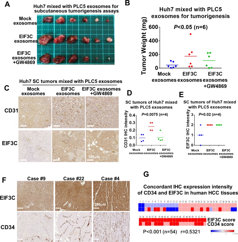
Figure 3. Various exosomes isolated from PLC5 mixed with Huh7 cells enhanced HCC angiogenesis and tumorigenesis.
(A) Subcutaneous tumorigenesis assays of Huh7 cells mixed with exosomes isolated from mock, EIF3C expression and EIF3C expression co-treated with GW4869. (B) Tumor weight and summary of subcutaneous tumorigenesis assays of Huh7 cells mixed with PLC5 exosomes isolated from mock, EIF3C expression and EIF3C expression co-treated with GW4869 (ANOVA summary). (C) Representative IHC staining of CD31 and EIF3C expression in PLC5 exosomes-enhanced subcutaneous Huh7 tumors with and without EIF3C expression and co-treatment of GW4869. (D) PLC5/EI3C exosomes enhanced CD31 expression is suppressed in compared to GW4869- treated PLC5/EIF3C exosomes-mediated Huh7 SC tumors (ANOVA summary). (E) Expression of EIF3C in PLC5/EIF3C-mediated Huh7 subcutaneous tumors showed no difference in compared to with and without treatments of GW4869 (ANOVA summary). (F) Representative IHC staining of EIF3C and angiogenic marker CD34 in human HCC tumors. (G) Heat map of concordant expression of CD34 angiogenic marker with EIF3C by IHC assays of HCC patients.

