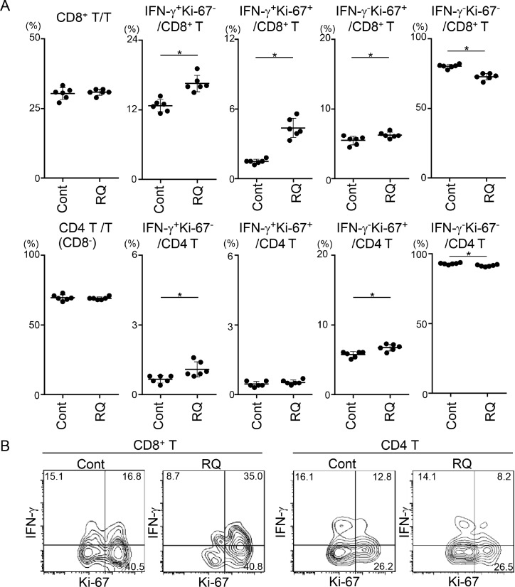
Figure 5. Resiquimod enhances activation of CD8+ T cells in RLNs and the TME.
RLNs (A) and tumor masses (B) from SCCVII-inoculated mice treated with control reagents or resiquimod at days 0, 3, 7, and 10 were obtained at 12 days. Tumor masses from five to six mice were combined, and TIL fractions were isolated. RLN cells and TILs were stimulated and stained with Brilliant Violet 510-anti-CD45, APC-eFluor780-anti-CD3, PerCP-Cy5.5-anti-CD8, APC-anti-IFN-γ, FITC-anti-Ki-67 mAbs or appropriate fluorochrome-conjugated control Abs. Stained cells were analyzed by flow cytometry. (A) Electric gates were placed on CD3+, CD3+CD8+, or CD3+CD8- (as CD4+ T) lymphocytes, and then the percentages of the indicated fractions were analyzed. Data shown are representative from two independent experiments with similar results. The bars show the mean values ± SD from each group of six mice. *Statistically different (p < 0.05). (B) An electronic gate was placed on CD45+CD3+CD8+ or CD45+CD3+CD8- (as CD4+ T) lymphocytes, and the expression levels of Ki-67 and IFN-γ are shown as contour plots. Data are representative from two independent experiments with similar results.

