Figure 3.
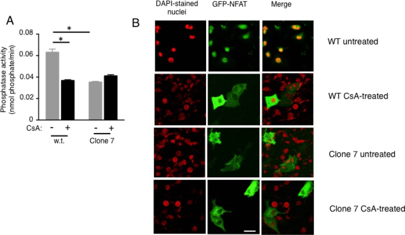
Calcineurin activity and NFAT nuclear localization are reduced in SELENOK deficient melanoma cells (A) Cells were incubated for 2 h with DMSO vehicle control or the calcineurin inhibitor, cyclosporin A (2 µM), and then cell lysates prepared normalized to total protein. The calcineurin substrate, RII phosphopeptide, was added to lysates and colorimetry used to detect production of phosphate. Means of replicates (N = 3) were compared using a student’s t-test and expressed as mean + SEM with *p < 0.05. All experiments are representative of a minimum of two independent experiments. (B) Using the same cyclosporine A conditions, GFP-NFAT localization was evaluated using confocal microscopy and results showed that SELENOK deficient Clone 7 cells had accumulation NFAT in the cytosol similar to cells treated with Cyclosporin A. Note that DAPI staining of nuclei was recolored from blue to red to allow merged (yellow) to be more apparent. Scalebar represents 5 µm.

