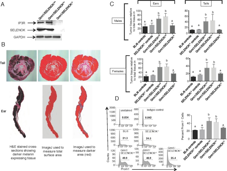
Figure 8. SELENOK deficiency reduces melanoma tumor growth on tails and ears.
(A) Western blot analysis of splenocytes, which express relatively high levels of SELENOK and IP3R, confirm that complete knockout of SELENOK leads to reduced IP3R in Grm1/SELENOK–/– mice. (B) Male and female littermates at 4 months of age with genotypes of Grm1/SELENOK+/+, Grm1/SELENOK+/–, and Grm1/SELENOK–/– were compared for levels of melanin positive tissue relative to total tissue in tails and ears. (C) Cross-sections of tails and ears were H&E stained and evaluated using an ImageJ color threshold approach as described in the Methods section. The dark melanin area relative to total area of tissue was compared for all three littermate genotypes. We also included two control groups expected to form no tumors (C57BL/6 and SELENOK–/–) due to a lack of the Grm1 transgene in melanocytes, which allowed assessment of melanin content in the absence of melanoma. Results showed that Grm1/SELENOK–/– mice had significantly reduced tumor growth in tails and ears, while Grm1/SELENOK+/+ and Grm1/SELENOK+/– mice were similar in progression of melanoma. Similar results were found with males and females. (D) Cells dissociated from were analyzed by flow cytometry for Prom1+ cancer stem cells and lower levels were found in Grm1/SELENOK–/– mice compared to Grm1/SELENOK+/+ and Grm1/SELENOK+/– littermates. Both sexes were included in each group, with representative images shown on the left and data graphed on the left (N = 3). A one-way ANOVA with Tukey post-test was used to analyze groups and means without a common letter differ, p < 0.05.

