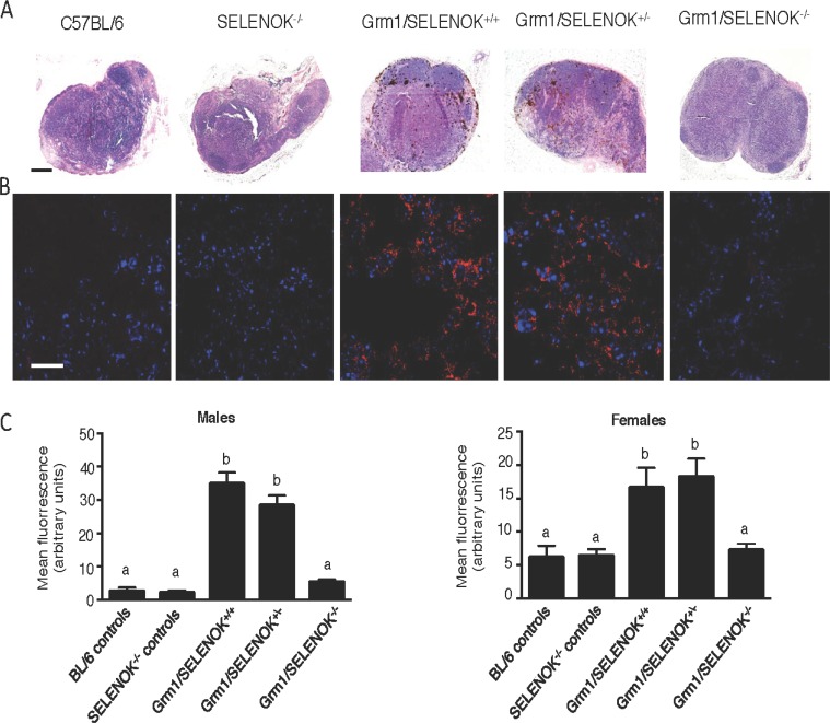
Figure 9. SELENOK deficiency reduces metastasis of melanoma to lymph nodes.
Male and female littermates at 4 months of age with genotypes of Grm1/SELENOK+/+, Grm1/SELENOK+/–, and Grm1/SELENOK–/– were analyzed for presence of melanoma cells in inguinal and axillary lymph nodes (N = 7/group). Non-tumor mice were included as controls (N = 3/group). (A) H&E stained lymph node sections were examined under low power (5×) for melanin, which appears as dark brown cells. Scalebar = 2 mm. (B) Immunofluorescence (40×) was used to detect the melanin specific antigen, Trp2 (red), in lymph node sections. DAPI (blue) was used to stain nuclei. Scalebar = 20 mm. (C) Trp-2 fluorescence was quantified using ImageJ with a minimum of 7 mice per group and 2 sections analyzed per mouse. A one-way ANOVA with Tukey post-test was used to analyze groups and means without a common letter differ, p < 0.05.

