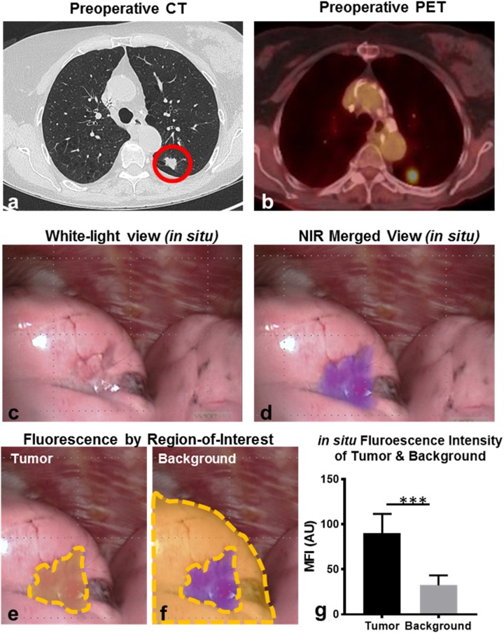
Figure 1. Pulmonary SCCs display in situ fluorescence during FR-IMI with OTL38.
Representative example: Subject 3 presented with a 1.7cm left upper lobe nodule by preoperative CT (a). Preoperative PET displayed an SUV of 8.7 (b). During FR-IMI the nodule was localized during standard thoracoscopic views (c), and displayed strong NIR signal during fluorescent imaging (d). Median fluorescent intensity (MFI) was determined for ROIs corresponding to tumor (e) and benign lung (background) (f). MFIs for ROIs were compared for SCCs (n=7) displaying in situ signal (g). Red circle-pulmonary SCC; yellow gates-ROIs measured by fluorescent analysis; ***p<0.001.

