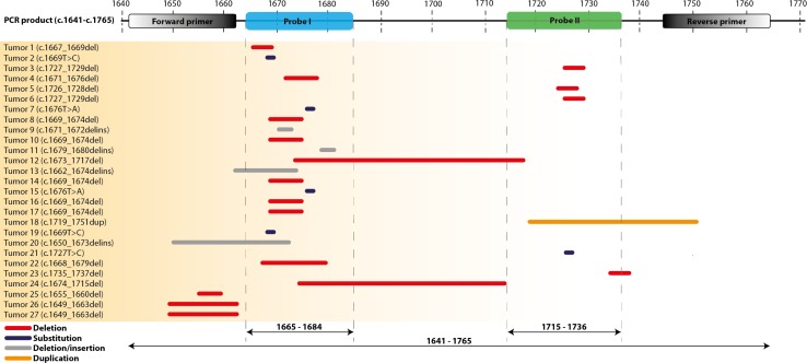
Figure 1. Location of KIT exon 11 mutations in GIST tumour samples as tested with Sanger sequencing or NGS.
The mutations are displayed relative to the actual position of the Forward and Reverse primers, the two probes (I and II) and PCR product. Type of mutations: red = deletion, grey = substitution, blue = deletion/insertion, orange = duplication.

