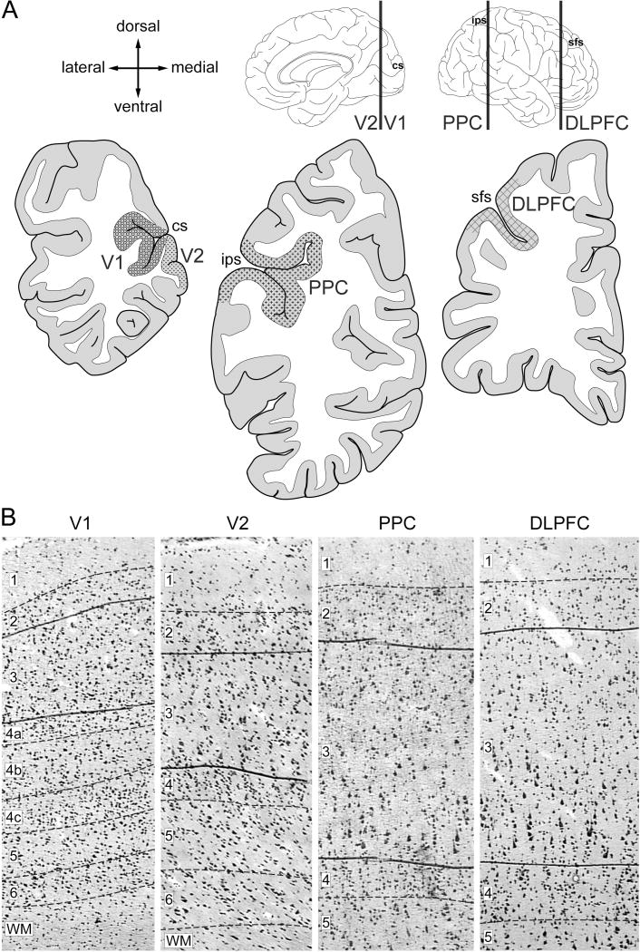
Figure 1. Sampling of cortical regions and layer 3.
(A) Schematic drawings of the medial and lateral surfaces of the right hemisphere of the human brain (top). Vertical lines indicate the approximate locations of the coronal sections (bottom). Locations of cortical regions of the vsWM network selected for sampling are shaded. Arrows labeled dorsal, medial, ventral, and lateral refer to the coronal sections (B) Representative Nissl-stained sections illustrating the laminar borders (dashed lines) and the borders as drawn of layer 3 for laser microdissection in each region. Numbers indicate cortical layers. V1, primary visual cortex; V2, association visual cortex; PPC, posterior parietal cortex; DLPFC, dorsolateral prefrontal cortex; sfs, superior frontal sulcus; ips, intraparietal sulcus; cs, calcarine sulcus.

