Fig. 5.
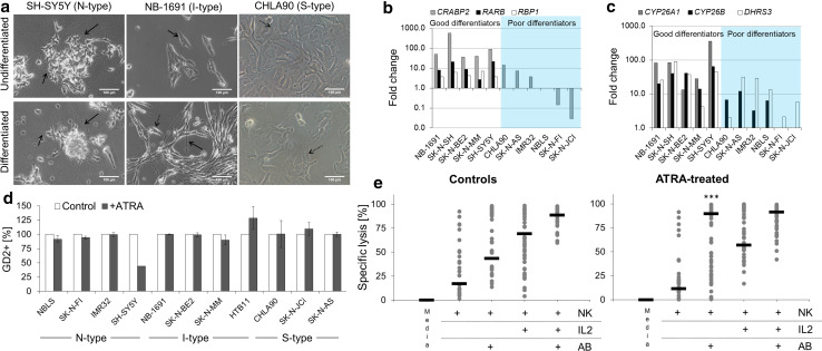
Effect of neuroblastoma differentiation on NK cell-mediated ADCC and ADCC. a After exposure to ATRA for 72 h, neurite outgrowth was present in N-type and I-type cells (solid arrows). No changes in morphology were noted in some I-type cells and most S-type cells (dashed arrows). b, c Differential gene expression of ATRA-binding proteins (b) and enzymes involved in ATRA metabolism (c) after differentiation of neuroblastoma cells. d Flow cytometry analysis of GD2. For all but two cell lines (SK-N-SH and SH-SY5Y), the percentage of GD2-expressing cells after ATRA-induced differentiation was unchanged, compared with that in untreated cells. e ADCC assays using 2 allogeneic donors against 10 neuroblastoma cell lines (listed in Supplementary Table 1) were conducted. Each dot refers to the average specific lysis per cell line for the respective experimental condition. Statistically significantly (***) higher median specific lysis (black bar) by resting NK cells and Hu14.18K322A was noted after differentiation with ATRA

