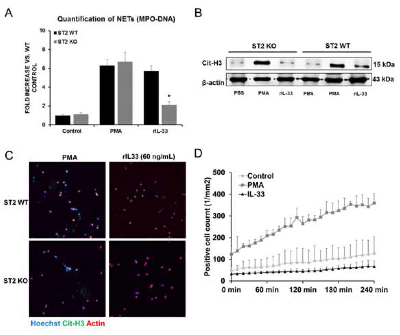
Fig. 4. NET formation is induced by IL-33 via ST2 signaling pathway.
(A) Quantification of MPO-DNA complexes in ST2 KO neutrophil media with rIL-33 treatment demonstrated a significant decrease (*P<0.05) compared to WT neutrophil media after stimulation. (B) ST2 WT neutrophils treated with rIL-33 exhibited increased cit-H3 compared to untreated neutrophils whereas ST2 KO neutrophils failed to citrullinate histone H3 in response to stimulation of rIL-33. (C) Confocal microscopy images revealed no NETs formation in ST2 KO neutrophils after rIL-33 stimulation. Green: IL-33; red: actin; blue: DAPI. (D) rIL-33 stimulation failed to induce NETs in ST2 KO neutrophils when quantified by Incucytes® intravital confocal microscopy.

