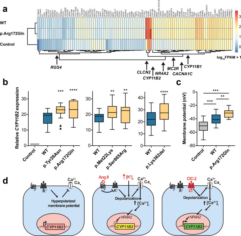
Figure 4. ClC-2 Increases Aldosterone Synthase Expression in H295R cells.
(a) RNA sequencing of H295R cells transfected with CLCN2 (WT or p.Arg172Gln), and vector control (heatmap). FPKM, fragments per kilobase of transcript per million fragments mapped. CLCN2 and CYP11B2 show the largest increase in expression versus control. Genes involved in adrenal function or calcium pathway are highlighted. (b) Relative expression levels of CYP11B2 (box, interquartile range; whiskers, 1.5× interquartile range; line, median) in the H295R cell line after transfection with empty vector (control), CLCN2WT (blue), and CLCN2MUT (yellow). Parallel transfections and real-time PCRs were performed in each group. CYP11B2 expression significantly increases after transfection of CLCN2MUT (see Supplementary Table 8 for statistical analysis). (c) Resting membrane potential (plots as in b) of HAC15 cells stably expressing CLCN2 (WT or p.Arg172Gln) and untransfected controls. WT and p.Arg172Gln cause significant depolarization versus control, and p.Arg172Gln causes significant depolarization versus WT (see Supplementary Table 8 for statistical analysis). (d) Model of ClC-2 function in human adrenal glomerulosa. Resting cells are hyperpolarized. Angiotensin II (AngII) and hyperkalemia cause depolarization, activation of voltage-dependent calcium channels, calcium influx, and increased CYP11B2 expression via the transcription factor NR4A2 (NURR1). ClC-2MUT causes increased CYP11B2 expression by membrane depolarization via increased Cl− efflux. **, p<0.01; ***, p<0.001; ****, p<0.0001.

