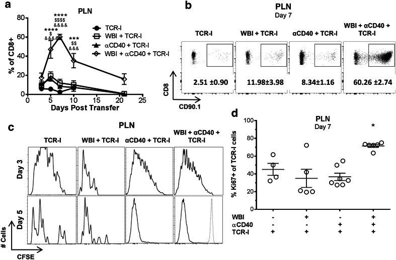
Fig. 3.
Dual conditioning prolongs donor T cell proliferation and increases accumulation in the PLN. a Percentage of CD90.1 + or TetI + TCR-I cells accumulating in the PLN over time. N = 6–18/group (days 3–10), 2–3/group (days 14, 21): WBI + αCD40 + TCR-I vs. TCR-I (asterisks), vs. WBI + TCR-I (dollar symbol), vs. αCD40 + TCR-I (ampersand); 1 digit p < 0.05, 2 digits p < 0.01, 3 digits p < 0.001, 4 digits p < 0.0001 by two-way ANOVA with Bonferroni corrections. b Representative flow cytometry panels from a. Numbers indicate the mean percentage +/− SEM of CD8+ cells. c CFSE staining of CD90.1 + TCR-I cells in the PLN 3 and 5 days post treatment. d Ki67 staining of CD90.1 + TCR-I cells in the PLN 7 days post treatment. N = 4–6/group; WBI + αCD40 + TCR-I vs. all other groups, **p < 0.01 by one-way ANOVA with Dunnett’s post-test

