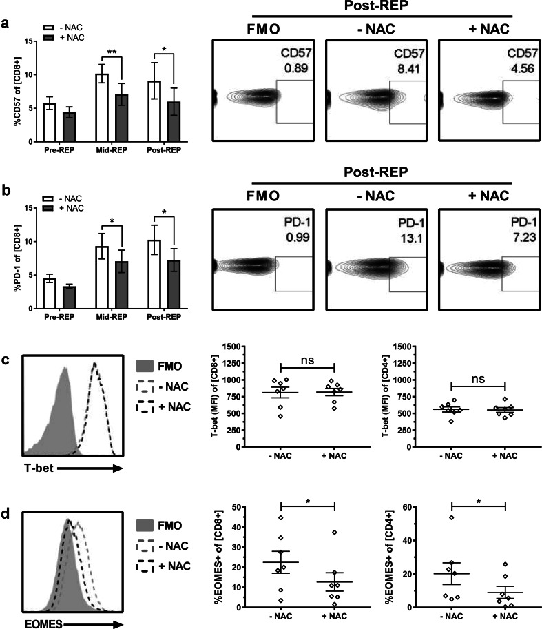
Fig. 3.
NAC impedes expression of senescence and exhaustion markers during rapid expansion. After 10 days in culture, TIL1383I TCR-transduced T cells were rapid expanded for an additional 10 days (± 2 mM NAC). Samples were cryopreserved before (Pre), on day 5 (Mid), and at the conclusion (Post) of the REP. Samples were subsequently analyzed for expression of a CD57 or b PD-1. Right panels display representative contour plots of post-REP cells. Left panels show quantification (mean ± SEM) of each indicated marker in CD34+ CD8+ cells over the course of the REP. n = 4 (2 melanoma patients, 2 healthy controls); *p < 0.05; **p < 0.01. TIL1383I TCR-transduced T cells rapidly expanded (± 2 mM NAC) were analyzed for the expression of c T-bet and d EOMES in CD34+CD8+-gated cells. Left panels display representative histogram overlay. Right panels are quantification (mean ± SEM) of n = 7. *p < 0.05; ns not significant

