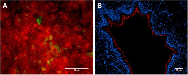
FIGURE 2.

Primary porcine respiratory epithelial cell models to study host–pathobiont interactions in the respiratory tract. Immunofluorescence microscopy analysis of (A) primary porcine bronchial epithelial cells under air–liquid-interface (ALI) conditions after 3 weeks of differentiation and (B) a precision-cut lung slice (PCLS). Ciliated cells were stained by β-tubulin antibody (shown in red, A + B), mucin-producing cells were visualized by mucin 5-AC antibody (shown in green, A), and nuclei were stained by DAPI (shown in blue, B). Bars represent 50 μm.
