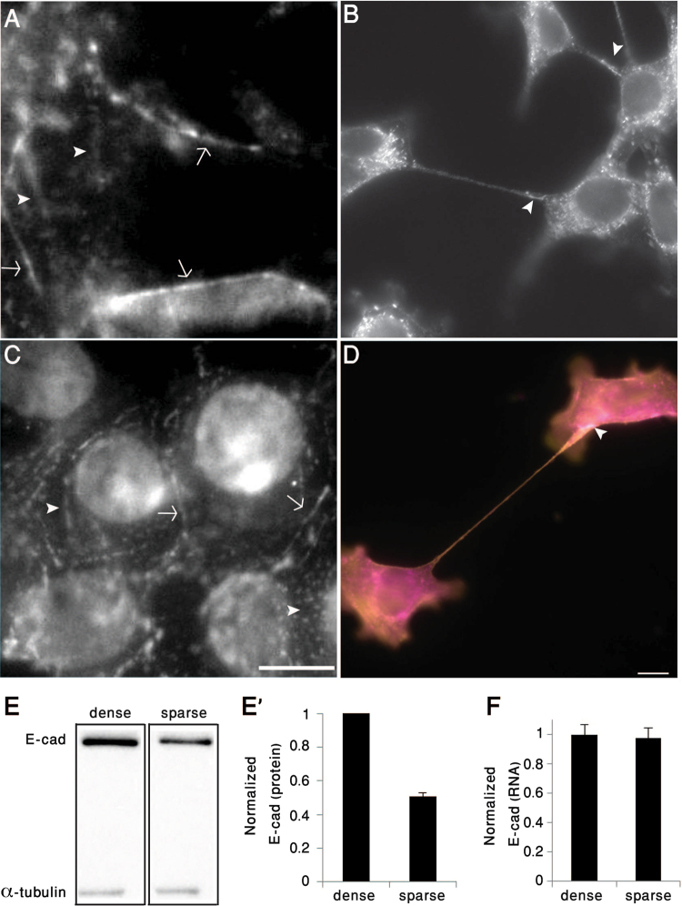
Figure 3.
Tether-mediated cell-cell adhesions are anchored via adherens-type junctions. (A,C) Densely-plated 4T1 cells form E-cadherin- (A) and β-catenin-rich (C) adherens junctions, located either along the sub-apical region (arrows), or in multiple dorsal patches (arrowheads). (B,D) Sparsely plated 4T1 cells form structurally and molecularly polarized tethers that are attached to the neighboring cell via E-cadherin (B), co-localized actin (magenta) and β-catenin (yellow) (D). Arrowhead in Panel D denotes the tether adhesion area. Scale bar: 10 μm. (E,E’) Immunoblotting and corresponding histogram depicting E-cadherin levels in sparsely- (5.6*103/cm2) and densely-plated (8.9*104/cm2) 4T1 cells. The normalized levels of E-cadherin (relative to α-tubulin) are shown in E’. Blotting Lanes of Fig. 3E were extracted from the same gel (full blotting of the gel is shown in Supplementary). (F) Comparison of E-cadherin mRNA levels in densely- and sparsely-plated 4T1 cells normalized to HPRT1 and GUSB genes. The results are based on two independent experiments.

