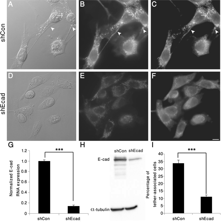
Figure 4.
E-cadherin is essential for tether formation. 4T1 cells, stably knocked down for E-cadherin (shEcad), and control cells (ShCon) were imaged by DIC (A,D) and double-labelled for E-cadherin (B,E) and β-catenin (C,F). Note that tether formation and E-cadherin-rich adhesions are markedly reduced in the knocked-down cells. (G) QRT–PCR analysis, pointing to an over 85% reduction in E-cadherin mRNA levels in shEcad compared to shCon 4T1cells, obtained in 3 independent experiments. (H) Western blotting revealed an ~83% decrease of E-cadherin protein levels in 4T1-shEcad, compared to 4T1-shCon. (I) Comparison of the percentage of tether-associated cells in shEcad and shCon 4T1cells, pointing to an ~68% reduction in the knocked-down cells, compared to controls. ***p < 0.001. Scale bar: 10 μm. n = 11 different images (containing around 800–900 cells in total for each group), obtained in two independent experiments.

