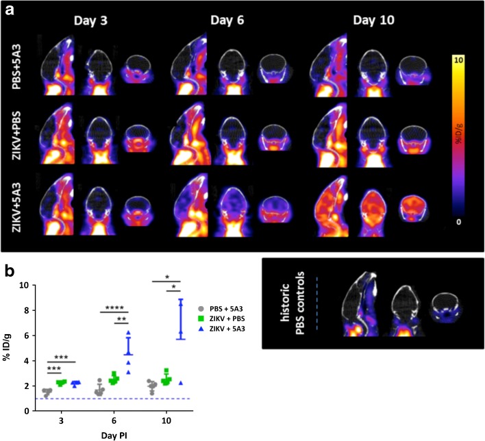
Fig. 2.
In vivo PET imaging can detect ZIKV-related neuroinflammation as early as day 3 post-infection (PI). Mice were separated into three treatment groups, PBS + 5A3, ZIKV + PBS, and ZIKV + 5A3 and were IV injected with [18F]DPA-714 at 3, 6, and 10 days PI to quantitatively assess brain neuroinflammation in terms of percent injected dose per gram tissue (%ID/g). a Representative PET/CT mouse brain images (sagittal, coronal, and transverse, from left to right) for each treatment group at days 3, 6, and 10 PI. Representative historic PBS control mouse brain images at bottom right. b Symbols, line, and error bars represent the individual mice, group mean, and standard deviation, respectively. The dotted line represents the mean [18F]DPA-714 binding value in brains from PBS-treated mice (~ 1.06 ± 0.21 %ID/g). *p < 0.05; **p < 0.01; ***p < 0.005; ****p < 0.001; one-way analysis of variance (ANOVA).

