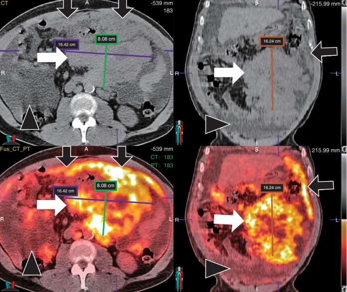
Fig. 6. Axial and coronal CT (upper panels) and fused FDG-PET/CT (lower panels) of a patient with new onset chylous ascites with fluid cytology consistent with B cell non-Hodgkin lymphoma.
Images show significant FDG-avid disease in the abdomen, including a heterogeneous necrotic mesenteric mass (white arrows), omental caking (black arrows), ascites (black arrowheads) and mesenteric and retroperitoneal lymphadenopathy.

