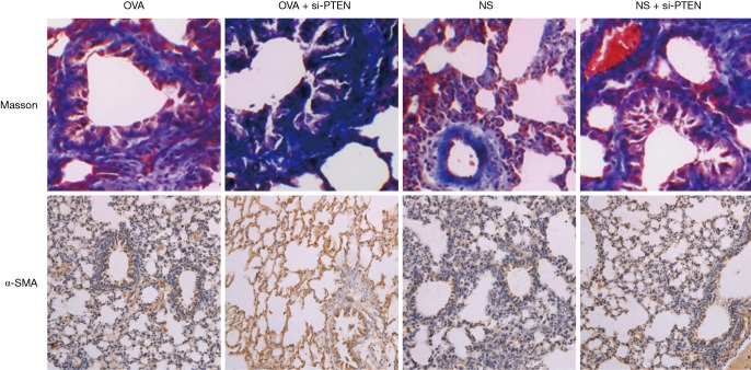
Figure 1.
Masson staining and α-SMA staining of lung tissue sections in the mice of each group (×200). Notes: Masson staining showed that collagen fibers appeared blue and muscle fibers appeared red. When immunohistochemistry was used to detect α-SMA, a brown appearance indicated a positive expression, and blue indicated negative expression. α-SMA, alpha smooth muscle actin; OVA, ovalbumin; NS, normal saline; si, small interference RNA; PTEN, phosphatase and tensin homolog.

