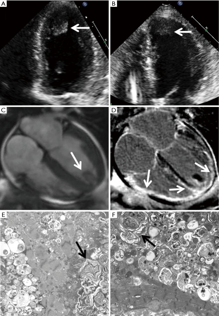
Figure 1.
HCQ-induced cardiomyopathy in twin sisters. The TTEs of the two sisters (A,B) show similar manifestation of LV hypertrophy with severely depressed systolic function and a large apical thrombus (arrow). A cardiac magnetic resonance was performed on the first sister and showed LV hypertrophy with severely decreased biventricular function (C) and confirmed the apical thrombus (arrow). Late gadolinium enhancement imaging showed extensive epicardial to mid myocardial enhancement involving the entire sub-basal mid to apical LV and the right ventricle (D, arrows). Electron microscopy of myocardial biopsies in two sisters (E,F) demonstrate myocardium with degenerative changes and numerous lamellar body inclusions (arrows). HCQ, hydroxychloroquine; TTE, transthoracic echocardiogram; LV, left ventricular.

