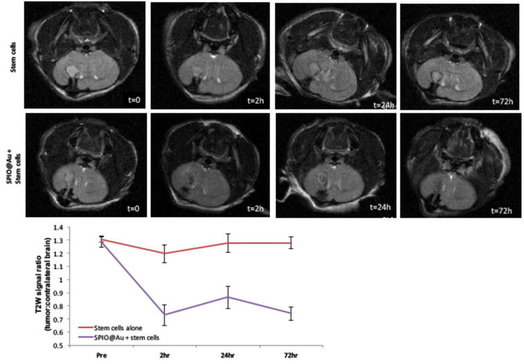
Figure 4.

Representative T2-weighted MR images of the mouse brain at various times (t = 0, 2h, 24h, and 72 h) after intra-carotid artery injection of SPIO@Au-loaded MSCs or unlabeled MSCs (top). Quantification of the T2-weighted signal of the tumor against that of the contralateral brain (bottom).
