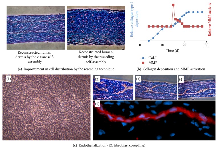
Figure 4.
Reseeding self-assembly technique. (a) Photographs of slices of tissue reconstructed by the classic self-assembly technique (left) or the reseeding self-assembly technique (right), stained with Masson's trichrome. Cells are in red/purple and ECM in blue. (b) Graph summarizing the balance between ECM production and its degradation during the production of a stroma by the self-assembly reseeding technique. Collagen deposition was measured by immunoblotting extracts from tissues produced until the indicated time. MMP activity was measured using tissue culture medium harvested at the indicated time using a fluorometric test. (c) (1) Phase contrast photograph of a coculture of endothelial cells and dermal fibroblasts (14 days after seeding). (2), (3), and (4) Photographs of slices of tissue reconstructed by the reseeding self-assembly technique, stained with Masson's trichrome. Three-dimensional capillary-like structures are clearly visible. (5) Photograph of a slice of tissue produced by the reseeding self-assembly technique immunostained using antibodies raised against CD-31.

