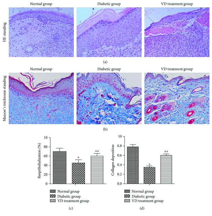
Figure 7.
Effects of vitamin D on the epithelialization and collagen deposition in ulcerative tissues at D14 after treatment. (a) H&E staining of sections showed better dermal reepithelialization on the diabetic wounds in the VD treatment group compared with the diabetic group. (b) Collagen deposition assessed by Masson's trichrome staining. (c, d) Statistical reepithelialization and thickness of collagen deposition of wounds by computer-assisted morphometric analysis. Data are represented as means ± SD. ∗p < 0.05, compared with the normal group. ∗∗p < 0.05, compared with the diabetic group.

