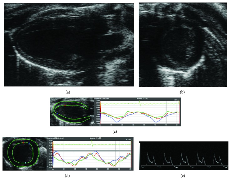
Figure 1.
Cardiac ultrasound scan. (a) Parasternal long axis view of the left ventricle. (b) Parasternal short axis view of the left ventricle. (c) Analysis of the longitudinal and radial strain/strain rate. (d) Analysis of the circumferential and radial strain/strain rate. (e) Parasternal 4-chamber view with PW-Doppler for the evaluation of E/A parameter.

